Professor of Biology at St. Olaf College Regents Hall of Natural Science Room 228
Gallery
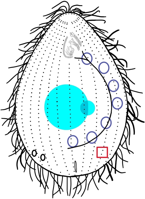
Diagram of a vegetative Tetrahymena Cell with clickable organelles.
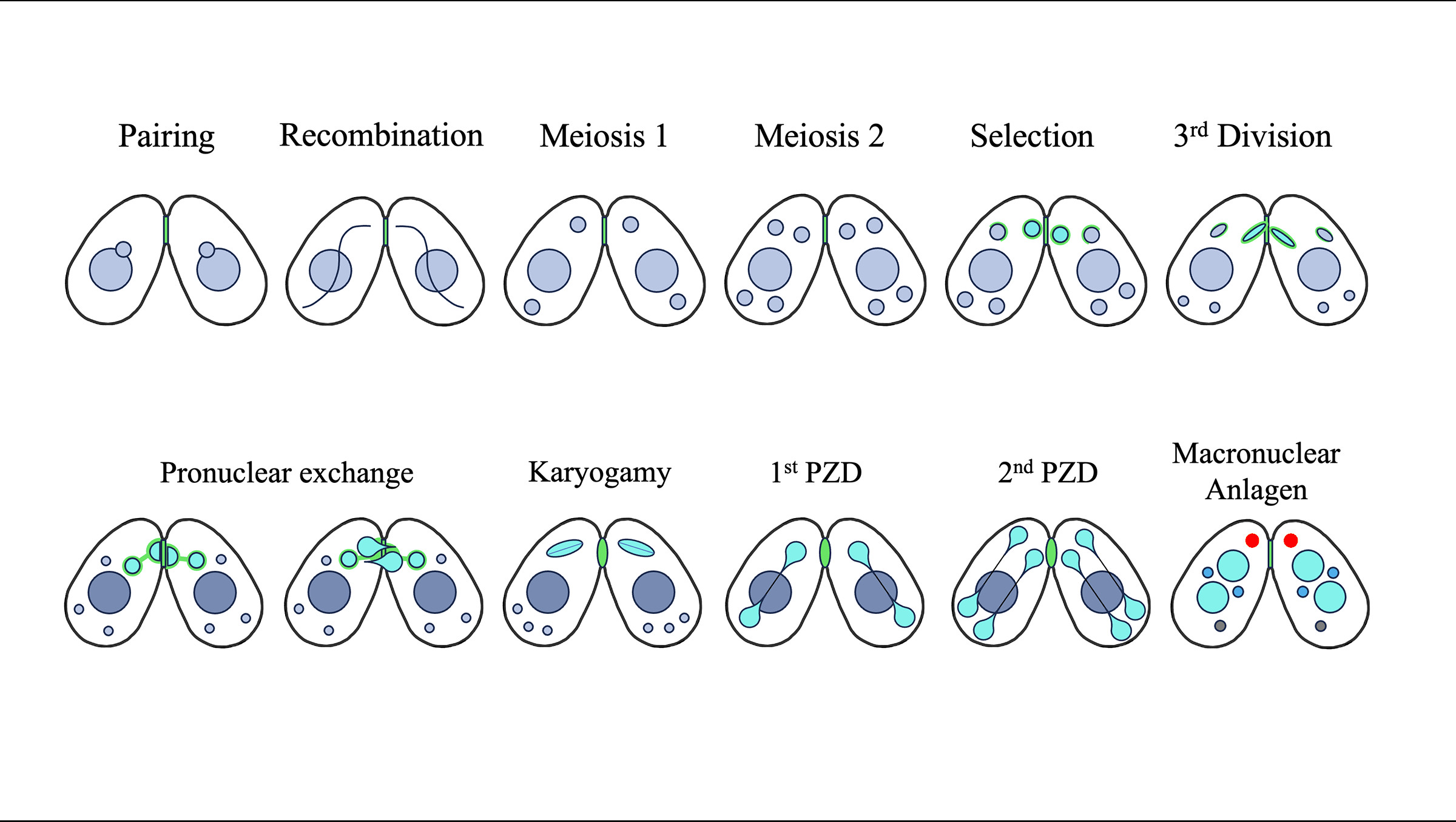
Diagram of the different phases of a Tetrahymena conjugation with clickable organelles.
Conjugation
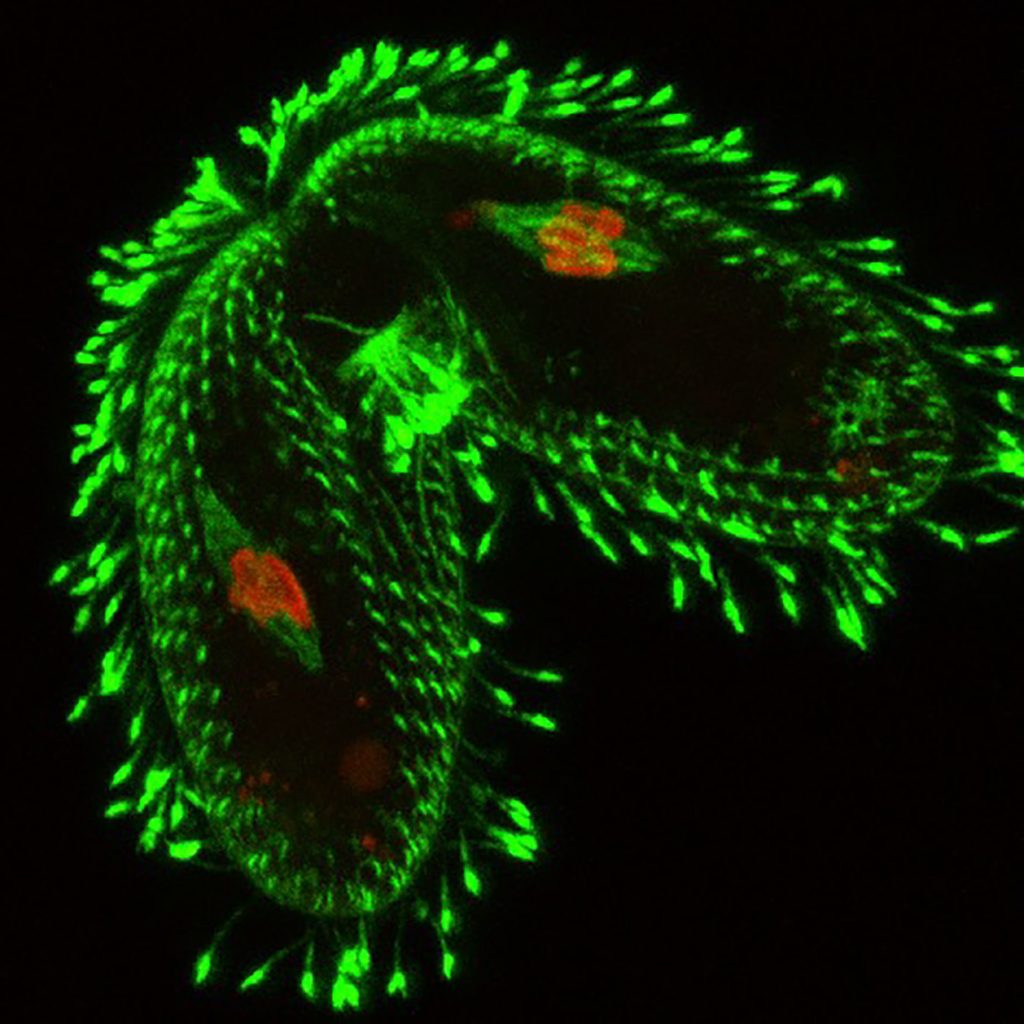
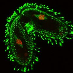
Animation of “Stomatogenesis” (new oral primordium) b y John J. Giannini. Open circles are un-ciliated basal bodies. Circlews with central dots are ciliated.Conjugal animation by John J. GianniniMating pair of Tetrahymena labeled to show cytoskeleton {antibody to acetylated alpha tubulin (green)} and micronuclei chromosomes {antibody to phosphorylated Histone H1 (red)}.
Vegetative division.
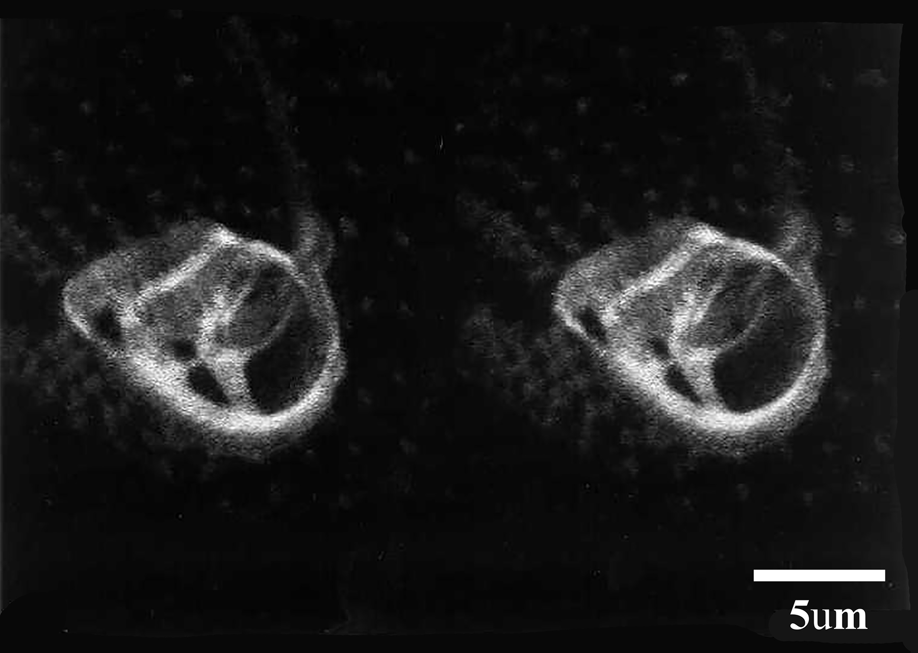
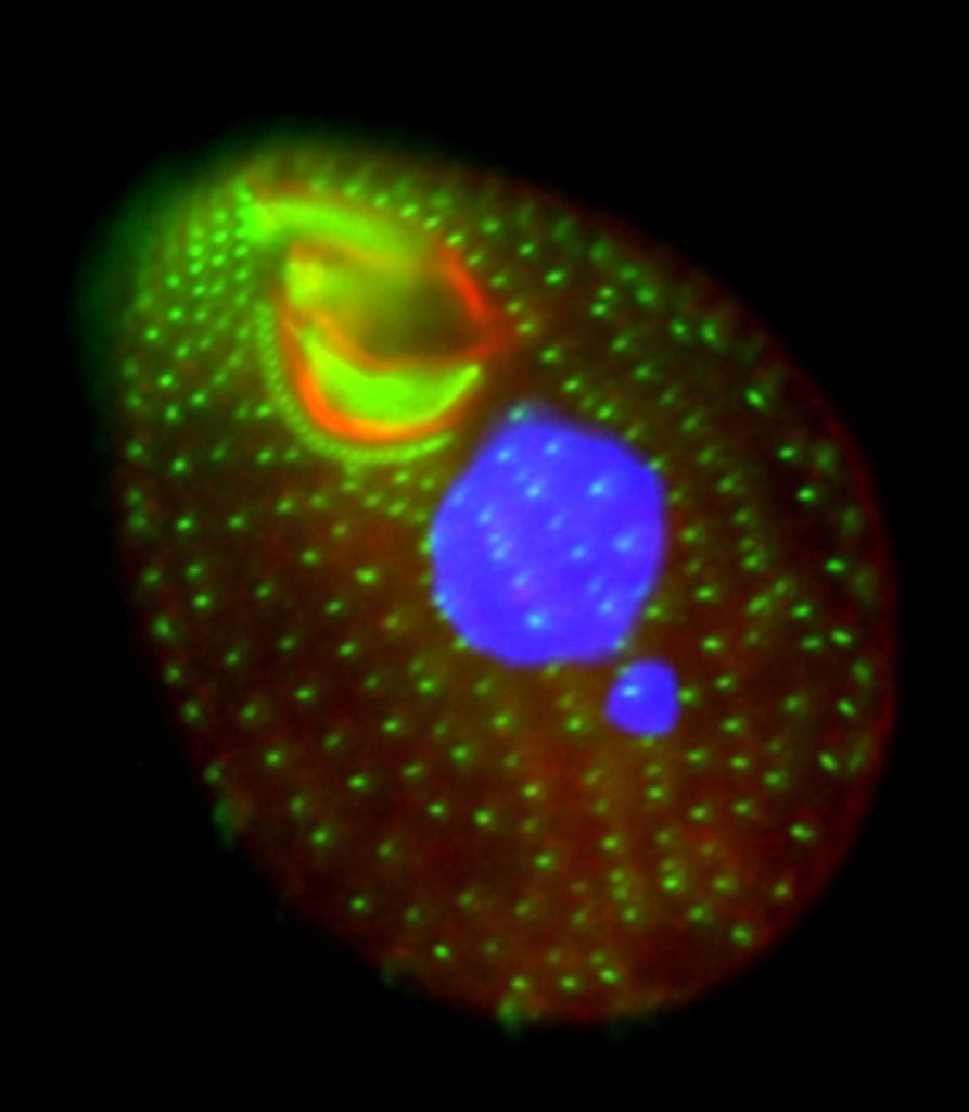
Stereo-Stereo pair of confocal micrographs of the Tetrahymena oral apparatus produced by double-staining with mAb 4F9 and mAb 11C7. The latter antibody is directed against tetrin 4, one of the oral filament proteins (see Honts and Williams, 1990). Unpublished photograph courtesy of Dr. N.E. Williams.Tetrahymena labeled with antibody to centrin (green) and an oral filament protein (red). Nuclei are labeled BLUE with DAPI. Conventional fluorescence microscopy.